Kidney Cancer Staging
From Kidney Cancer Resource
Contents |
Overview
Determining the extent of the spread or the stage of the cancer requires a number of tests. Staging tests include X-rays, computerized tomography (CT) scans, ultrasonography or magnetic resonance imaging (MRI). Other tests include an intravenous pyelogram (IVP) and arteriography. Intravenous pyelogram involves the injection of dye into a vein to help visualize the kidneys, ureters and bladder. If the patient has bone pain, recent bone fractures, or certain abnormalities on the blood tests, a bone scan is also recommended. Additional tests may be obtained as needed. Kidney cancer has the propensity to grow into the renal vein and vena cava. The portion of the cancer that extends into these veins is called “tumor thrombus.” Imaging studies help determine if tumor thrombus is present. There are no blood or urine tests that directly detect the presence of kidney tumors. Arteriography involves the injection of dye into the blood vessels supplying the kidney. Staging is ultimately confirmed by surgical removal of the cancer and exploration of the area adjacent to the kidney. The surgeon will often remove regional lymph nodes for examination under the microscope. Examination of both kidneys is essential to assure that one is working normally. Sometimes, more progressed stages of the disease can be determined by such tests without the need for surgery.
In 40% of patients, renal cell cancer will be limited to the kidney and is treated exclusively by surgery, which is curative 90% of the time. In the 60% of patients with renal cell cancer that has spread outside the kidney, the disease is generally not curable with surgery and other specialists, such as medical oncologists and possibly even radiation therapists, are involved with treatment.
Following completion of all diagnostic tests and surgery, a final "pathologic" stage and grade will be given. All new treatment information concerning renal cell cancer is categorized and discussed by the stage. Tumor grade is a subjective measure of how aggressive the tumor looks under the microscope; therefore, it is determined from a surgical specimen. Grade cannot be determined from radiographic imaging (CT scans, MRI, etc..), blood tests or urine tests. Grade usually ranges from 1 to 4 with higher numbers indicating a more aggressive tumor. Thus, higher grade implies a worse prognosis.
The following are simplified definitions of the various stages of kidney cancer. Click on each for a stage by stage overview of the most recent information available concerning the comprehensive treatment of renal cancer.
Stage I
The primary cancer is 7 centimeters (about 3 inches) or less and is limited to the kidney, with no spread to lymph nodes or distant sites.
For more details on Click Here
Stage II
The primary cancer is greater than 7 centimeters (about 3 inches) and is limited to the kidney, with no spread to lymph nodes or distant sites.
For more details on Click Here
Stage III
The primary cancer is less or greater than 7 centimeters (about 3 inches), but has spread to only a single regional lymph node. The primary tumor may have spread to the renal veins or vena cava (large vein returning blood to the heart located in the middle of the abdomen near the back), but has only spread directly and not out of the local area of the kidney.
For more details on Click Here
Stage IV
The cancer has spread to distant sites, invades directly beyond the local area or has more than one lymph node involved.
For more details on Click Here
Recurrent Renal Cell Cancer
Renal cell cancer has returned after primary treatment with surgery, chemotherapy, radiation or biological modifiers.
Clinical stage is based on radiographic imaging before surgery, whereas pathologic stage is based on the analysis of surgically removed tissue. Staging the cancer helps predict prognosis and survival.
Physicians further denote the stage of a cancer according to a system developed by the American Joint Committee on Cancer (AJCC). This staging system includes the following criteria:
1.) the size or extent of the primary kidney tumor growth into the kidney or T stage
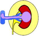
| Primary Tumor Stage (T stage) | Graphic Representation | Description |
| T1 | 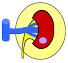
| Tumor is confined to the kidney (i.e. no penetration through the capsule) and is7 centimeters or less in greatest dimension |
| T2 | 
| Tumor is confined to the kidney (i.e. no penetration through the capsule) and is greater than 7 centimeters in greatest dimension |
| T3a | 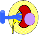
| Tumor penetrates through the kidney capsule into the surrounding fat or the adrenal gland, but not through Gerota’s fascia. |
| T3b or T3c | 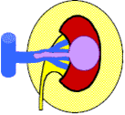
| Tumor extends into the renal vein or into the vena cava.
-T3b indicates that the tumor thrombus does not extend above the level of the chest diaphragm. -T3c indicates that the tumor thrombus extends above the level of the chest diaphragm |
| T4 | 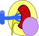
| Tumor penetrates through Gerota’s fascia. |
2.) the status of lymph nodes near the kidney or N stage (in renal cell cancer the lymph nodes near the kidney are referred to as regional lymph nodes); and
Regional Lymph Nodes (N stage) Description
N0 No cancer in the lymph nodes
N1 Cancer in a single lymph node
N2 Cancer in more than one lymph node
3.) the presence or absence of cancer spread to distant sites (metastases) or M stage Distant Metastasis (M Stage) Description M0 No metastasis M1 Distant metastasis present
In general, cancers with higher T stage, lymph node metastasis (N stage) or distant metastasis (M stage) are associated with a worse prognosis and typically shorter survival periods.
References
|
Disclaimer
Kidney Cancer Resource (KCR) is not influenced by sponsors. The information contained herein is not intended as a substitute for the advice of an appropriately qualified and licensed physician or other licensed health care provider. The information provided here is for educational and information purposes only. Early accurate Diagnosis (Dx.) saves lives. Please check with a physician if you suspect you are ill, never ignore Symptoms. To help your health care specialist make an accurate Diagnosis please keep notes of dates, times and details of your Symptoms. We are not offering medical advice nor do we consider links, individuals or articles accessed through this site to be offering medical advice.
E&OE - Errors & Omissions Excepted
As much of the information posted on this Web Site for peoples convenience is of a medical or technical nature, and may be a matter of life or death the E&OE is a Disclaimer showing that to the best of our ability information is accurate and correctly written or transcribed. Before acting on information on this site you are responsible for checking it with your relevant medical team. We can not be held responsible for any Errors & Omissions made; nor for information on links and articles provided in good faith.
